Cause and presentation
This lesion is usually unilateral and affects several structures in the midbrain including:
| Structure damaged | Effect |
|---|---|
| substantia nigra | contralateral parkinsonism because its dopaminergic projections to the basal ganglia innervate the contralateral hemisphere motor field |
| corticospinal fibers | contralateral hemiparesis and typical upper motor neuron findings |
| corticobulbar tract | difficulty with contralateral lower facial muscles and hypoglossal nerve functions |
| oculomotor nerve fibers | ipsilateral oculomotor nerve palsy with a drooping eyelid and fixed wide pupil pointed down and out. This leads to diplopia |
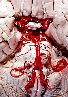
It is caused by midbrain infarction as a result of occlusion of the paramedian branches of the posterior cerebral artery or of basilar bifurcation perforating arteries.[1]
Weber's Syndrome has presented as a manifestation of decompression illness in a recreational scuba diver
58 一位65歲男性,突發性頭暈、嘔吐、口齒不清且吞嚥困難。身體檢查發現左側瞳孔較小且眼瞼下垂。請問最可能的病因是:
左側後交通動脈之動脈瘤破裂(posterior communicating artery aneurysm rupture)
左側後下小腦動脈阻塞(posterior inferior cerebellar artery occlusion)
左側豆紋狀動脈阻塞(lenticulostriate artery occlusion)
左側中大腦動脈阻塞(middle cerebral artery occlusion)
answer B PICA
this case is refering to pica obstruction
resulting in
PICA / wallenberg syndrome
horner syndrome
http://en.wikipedia.org/wiki/PICA_syndrome
The clinical features of Horner's syndrome can be remembered using the mnemonic, "Horny PAMELa" for Ptosis, Anhidrosis, Miosis, Enophthalmos and Loss of ciliospinal reflex.
ysfunction Effects vestibular nuclei vestibular system: vomiting, vertigo, nystagmus, inferior cerebellar peduncle Ipsilateral cerebellar signs including ataxia, dysmetria (past pointing), dysdiadokokinesia central tegmental tract palatal myoclonus lateral spinothalamic tract contralateral deficits in pain and temperature sensation from body (limbs and torso) spinal trigeminal nucleus & tract ipsilateral loss of pain, and temperature sensation from face nucleus ambiguus - (which affects vagus nerve and glossopharyngeal nerve - localizing lesion (all other deficits are present in lateral pontine syndrome as well) ipsilateral laryngeal, pharyngeal, and palatal hemiparalysis: dysphagia,hoarseness, diminished gag reflex (efferent limb - CN.X) descending sympathetic fibers ipsilateral Horner's syndrome (ptosis, miosis, & anhydrosis)
(middle cerebral artery occlusion)
The MCA is by far the largest cerebral artery and is the vessel most commonly affected by cerebrovascular accident (CVA). The MCA supplies most of the outer convex brain surface, nearly all the basal ganglia, and the posterior and anterior internal capsules. Infarcts that occur within the vast distribution of this vessel lead to diverse neurologic sequelae. Understanding these neurologic deficits and their correlation to specific MCA territories has long been researched.

Branches
The middle cerebral artery can be classified into 4 parts:[2]
- M1: The sphenoidal segment, so named due to its origin and loose lateral tracking of the sphenoid bone. Although known also as the horizontalsegment, this may be misleading since the segment may descend, remain flat, or extend posteriorly the anterior (dorsad) in different individuals. The M1 segment perforates the brain with numerous anterolateral central (lateral lenticulostriate) arteries, which irrigate the basal ganglia.
- M2: Extending anteriorly on the insula, this segment in known as the insular segment. It is also known as the Sylvian segment when the opercular segments are included. The MCA branches may bifurcate or sometimes trifurcate into trunks in this segment which then extend into branches that terminate towards the cortex.
- M3: The opercular segments and extends laterally exteriorly from the insula towards the cortex. This segment is sometimes grouped as part of M2.
- M4: These finer terminal or cortical segments irrigate the cortex. They begin at the external of the Sylvian fissure and extend distally away on the cortex of the brain.
- Contralateral hemiplegia affecting face, arm, and leg (lesser).
- Homonymous hemianopia - Ipsilateral head/eye deviation.
- If on left: global aphasia.
- Bilateral occlusion of Anterior Cerebral Arteries at their stems results in infarction of the anteromedial surface of the cerebral hemispheres:
- Paraplegia affecting lower extremities and sparing face/hands.
- Incontinence
- Abulic and motor aphasia
- Frontal lobe Symptoms: personality change, contralateral grasp reflex.
- Unilateral occlusion (distal to Ant. Comm. origin) of Anterior Cerebral Artery produces contralateral sensorimotor deficits mainly involving the lower extremity with sparing of face and hands (think of the humunculus).
Pathology
Aneurysms of the posterior communicating artery are the second most common circle of Willis aneurysm[1] (the most common are anterior communicating arteryaneurysms) and can lead to oculomotor nerve palsy

No comments:
Post a Comment